| |
|
|
Dr Rajesh Gajbhiye
Dr Rakhi Gajbhiye
Consultant gynecologists
Mauli Women’s Hospital,
Chhaoni,
Nagpur.
A 21 years old girl came with swelling in vulva since 18
years. She was a case of bilateral vulval hemangioma. The
swelling increases on standing and coughing and is reduced
on lying down position. She was operated for right vulval
hemangioma in 1990 at railway hospital. She was again
operated for left vulval hemangioma in 1993.It was said to
be cavernous hemangioma. She had a residual right hemangioma
for which she was given injection of 3% Sodium tetradeacyl
sulphate under short GA. this injection was repeated every
six weeks for a year but there was no relief. In 1998 again
she was treated with local sclerosant injections fro one
year but had no relief. She had undergone surgery for
vesical calculus in January2008. CT scan report was vesical
calculus with multiple phleboliths in pelvis. On local
examination she had a swelling on right labia majora which
on standing position was about 6x7 cm. It was reducible and
soft on palpation. (Fig 1)
She was investigated and planned for excision of hemangioma.
Her blood works were with in normal limits especially
platelet count and coagulation profile was normal. Colour
Doppler was done which showed no feeder vessel from large
vessel and slow venous flow. Her excision of hemangioma was
done. There were cavernous spaces. Excision of spaces was
done till the periosteum of pubic bone. At places feeder
vessels were identified and hemostasis achieved. There were
phelboliths scattered in that mass which were removed. Drain
was kept and incision was closed for two days patient was
fine but when the drain was being removed she had profuse
bleeding. Immediately the pressure was given and three blood
transfusions were given. Injectable Tranexamic acid was
given round the clock. Subsequently the drain was removed.
There was a wound gaping which responded to dressing.
Patient is doing fine now but has mild swelling on left
side.
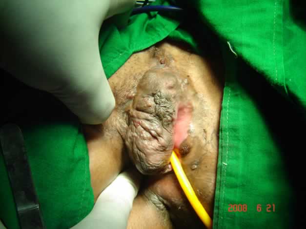
Fig 1: Cavernous Hemangioma
Discussion:
Kondi-Pafiti A et al in their clinicopathological study of
nine cases found one capillary hemangioma of the
endometrium, one capillary and one cavernous hemangioma of
the cervix, six vulva lesions (one capillary, and one
cavernous hemangioma and four angiokeratomas) and one
hemangiosarcoma of the mons pubis. Gupta et el in their
study of vascular tumors of female genital tract, found The
vascular tumors occurred most commonly in ovary (six),
followed by vulva (two), and one each in cervix and vagina.
Clinical diagnoses ranged from cystadenoma in ovarian tumors
to endocervical polyp in cervical tumor. Histologically, all
were benign vascular neoplasms, ranging from hemangioma
(five), lymphangioma (one), lymphangioma circumscriptum
(one) to angiomatosis (two) and arteriovenous malformation
(one). Klippel-Trenaunay-Weber syndrome is a sporadic
genetic syndrome characterized by localized hemangiomas,
venous varicosities, and asymmetric osseous hypertrophy of
the ipsilateral extremities. Most commonly seen in
association with hemangiomas, Kasabach-Merritt syndrome is
defined by the presence of thrombocytopenia and a
consumptive coagulopathy
Bava et al reported a case of a vulvar hemangioma
complicated by life-threatening hemorrhage not responsive to
corticosteroids therapy, which showed no signs of
spontaneous involution. The therapeutic approach consisted
of 3 steps of selective arterial embolization followed 5
months later by surgical excision and reconstruction of the
vulva. Selective embolization rarely is used in the
treatment of hemangiomas but may be required in cases of
intractable bleeding, severe heart failure, large and poorly
involuting hemangiomas, and as preparation for surgery
Vulvar venous malformations should be distinguished from vulvar varicosities, hematomas, soft-tissue neoplasms, and
other vascular anomalies. Doppler ultrasound, MRI, and
direct-injection venography are the most accurate diagnostic
modalities.
References
1.
Kondi-Pafiti A, Kairi-Vassilatou E, Spanidou-Carvouni
H, Kontogianni K, Dimopoulou K, Goula K. Vascular tumors of
the female genital tract: a clinicopathological study of
nine cases. Euro J Gynecol Oncol. 2003; 24(1):48-50.
2.
Bava GL, Dalmonte P, Oddone M, Rossi U.,
Life-threatening hemorrhage from a vulvar hemangioma., J
Pediatric Surg. 2002 Apr;37(4):E6.
3.
Kasabach-Merritt coagulopathy complicating
Klippel-Trenaunay-Weber syndrome in pregnancy. Obstet
Gynecol. 1995 May;85(5 Pt 2):831
4.
Gupta R, Singh S, Nigam S, Khurana N Int J Gynecol
Cancer. 2006 May-Jun;16(3):1195-200
5.
Neubert AG, Golden MA, Rose NC, Kasabach-Merritt
coagulopathy complicating Klippel-Trenaunay-Weber syndrome
in pregnancy. Obstet Gynecol. 1995 May;85(5 Pt 2):831-3
|
|